Anatomy and Physiology: All Parts, Big and Small
All Parts, Big and Small
To understand how a muscle contracts, it is helpful to think of those old Roman ships with the galley of slaves rowing the oars in unison. Each slave had only so much strength, but all their strength combined, not to mention the ability to row together in unison, gave the ship's oars incredible power.
A muscle not only has multiple units, but also needs that connect it with the rest of the body. Being irritable, muscles need to have a connection to motor neurons. All the need for ATP means the need for O2 and CO2 exchange, not to mention H2O, waste, and glucose transport. These needs all require the presence of blood, lymph, and the vessels to carry them. Nerves, blood vessels, and lymphatics are held in connective tissue around the muscles, tissues known as fascia.
Fascia Galore!
Fascia refers to connective tissue that surrounds muscle, which provides both protection and flexibility. It is helpful if you have ever cooked chicken and had a chance to handle both raw and cooked meat, then you'll know fascia. Meat is, after all, muscle! The act of cooking causes the fascia to break down, which makes the comparison of raw and cooked meat very useful. You might never be able to look at meat the same way again, but you are, after all, what you eat!
Superficial fascia is so called because it is the closest to the skin, being immediately deep to the skin (which is why it is also called subcutaneous fascia). If you have ever eaten cooked chicken you probably noticed that the skin comes off easily. On the other hand, removing the skin from raw chicken is not so easy! To do so you need to pull up on the skin and gently cut the superficial fascia that holds the skin to the muscle. This fascia breaks down during cooking, which is why it's so easy to take the skin off after cooking.
This layer of fascia also has adipose tissue (fat), and it is where most excess fat is stored when you exceed your ideal body weight. In addition to storing fat and water, this layer provides insulation from heat loss, a cushion-like protection from trauma, and a place for vessels and nerves in and out of muscles. Figure 8.1 shows a view of the fascia.

Figure 8.1Fascia appears at several levels in and around every muscle. (©2004 Pearson Education, Inc., publishing as Benjamin Cummings)
Since muscles come in groups, these groups are held together, with division to separate them by a deeper layer of fascia called, what else, deep fascia. This layer, by providing a division between muscles, helps the muscles to contract without interference. Each muscle in the group has its own layer of outer fascia called epimysium. Thus each muscle in the group has its outer epimysium, all of which is surrounded by deep fascia.
Fascicles
Have you ever noticed that meat that is overcooked tends to be stringy? That illustrates an interesting point—that muscles are made of many parallel fibers that contract in the same direction. Individual muscle cells are called myofibers, and these myofibers are arranged in groups of around 10 to 100, and these groups are called fascicles. Between these myofibers of every fascicle is a connective tissue called endomysium. Finally, around every fascicle is another layer of fascia called perimysium (peri meaning around, in this case around the fascicle). When you overcook meat the perimysium breaks down, thus highlighting the individual fascicles, making the meat look stringy!
Myofibers
This layer upon layer effect continues in each muscle cell or myofiber. In case you haven't noticed, myo means muscle; so when you read myofiber think muscle fiber. These cells have a few terms of their own. For instance, using another prefix, sarco-, which also means muscle, the cytoplasm becomes the sarcoplasm, and the E.R. (endoplasmic reticulum) becomes the S.R. (sarcoplasmic reticulum).
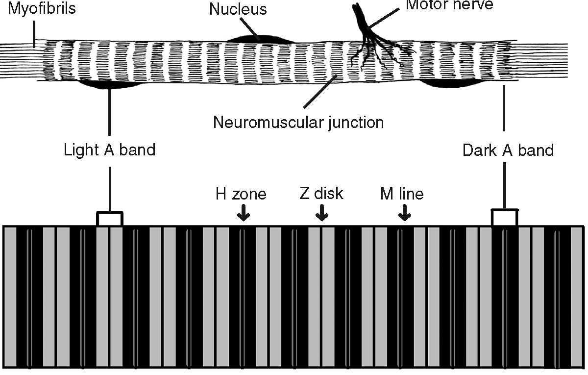
Figure 8.2Two views of a myofiber. (©2003 www.clipart.com and ©Michael J. Vieira Lazaroff)
Given the importance of the membrane in receiving the message to contract, the myofiber membrane becomes the sarcolemma. Not to be outdone, the neuron's cell membrane is called the neurilemma. The arrangement of proteins in a muscle cell is its crowning glory, and the thread-like proteins in the sarcoplasm are called myofibrils. These myofibrils form striations (see Figure 8.2) in skeletal and cardiac muscle of alternating myofilaments (thin and thick filaments), which I will explore when I discuss the unit of muscle contraction, the sarcomere.
Excerpted from The Complete Idiot's Guide to Anatomy and Physiology © 2004 by Michael J. Vieira Lazaroff. All rights reserved including the right of reproduction in whole or in part in any form. Used by arrangement with Alpha Books, a member of Penguin Group (USA) Inc.
To order this book direct from the publisher, visit the Penguin USA website or call 1-800-253-6476. You can also purchase this book at Amazon.com and Barnes & Noble.
Trending
Here are the facts and trivia that people are buzzing about.