Anatomy and Physiology: Stops Along the Way
Stops Along the Way
In order to understand the digestive system, it is important to think about the different organs and the varied roles they play (see Figure 14.1). First let's get to know their names. In terms of the organs of the digestive tract itself, if it's easier, think “My Pink Elephant Still Smells Like Rotten A,pples.” Whaaaat? Simple. Mouth, Pharynx, Esophagus, Stomach, Small Intestine, Large Intestine, Rectum, Anus. These amazing organs, despite the wonderfully gross things they do, still need help. The accessory organs (given their importance, it's a bit odd to call them that)—salivary glands, pancreas, liver, and gallbladder—will be discussed in more detail later.

Figure 14.1This view shows the overall layout of the major digestive organs, although some are hidden from view. (LifeART©1989-2001, Lippincott Williams & Wilkins)
What They Have in Common
Medical Records
Given our evolutionary past as organisms that absorbed food through our outer layer, the lining of our mouth to the lining of our intestines is all continuous with the outside of the body; this makes the inside of your stomach technically the outside of your body! The outside of our body protects itself by making the outer layer dead tissue, but given the need to do everything from producing enzymes to absorbing food, the inside of our digestive tract needs to be alive. For this reason, we risk bacterial invasion from both ends, and a normal, healthy person has bacteria in both the mouth and the large intestine.
Despite their differences, it's a good idea to find what all these organs have in common. The organization of our digestive tract in our basic body plan hasn't really advanced much beyond that of the earthworm (phylum Annelida); as such, it is hundreds of millions of years old. Our basic body plan is that of a tube within a tube. The inner tube is, of course, your digestive tract, but the outer tube is your larger hollow body cavity, which is divided into the thoracic and abdominopelvic cavities.
Given that every organ is made of many tissues, it is important to look at the histology, from the inside out, of the layers of the typical digestive organ. All these layers are illustrated in Figure 14.2. After all, as with people, they are more alike than they are different.
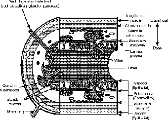
Figure 14.2This view of the small intestine illustrates all the major layers of every organ of the digestive tract.
The innermost layer is called the mucosa. The mucosa, which is, as you would imagine, a mucous membrane, varies most from organ to organ among all the layers. Just outside the mucosa is the lumen, or hollow cavity, of the organ. This lumen is where all the mechanical and chemical digestion happens. This type of digestion is called extracellular digestion (meaning outside the cell).
The cells of the mucosa produce enzymes that are released by exocytosis, and the food is digested outside the cells, which is literally outside the body. None of the food actually enters the body until it is absorbed by the mucosa. This layer is composed of epithelial tissue, which is avascular (without blood vessels). It is this layer that secretes the enzymes, as well as the chemical protection from those enzymes.
The submucosa, which is as the name implies under the mucosa, is, like all layers beneath epithelial layers, made of connective tissue. This is where blood vessels and lymphatics (see Cardiovascular and Lymphatic Circulation) can be found; the food absorbed by the mucosa is then absorbed into these vessels. Many of the glands of the mucosa also extend into this layer.
The Big Picture
Within each of the connective tissue layers are simple connections to other body systems. The capillaries connect the organs to the cardiovascular system. Hormones of the endocrine system also travel through these capillaries. Drainage and uptake of lipids are accomplished through vessels of the lymphatic system, known as lymphatics. Nerves from, of course, the nervous system can also be found here, otherwise our brain would have no idea what is happening down there!
Peristalsis, the movement of food along the digestive tract, is accomplished in the next layer, the muscularis externa. This layer consists of two separate layers of smooth muscle, a longitudinal layer, and a transverse, or circular, layer; peristalsis consists of alternating contractions of the two layers. The immense amount of churning in the stomach requires the existence of a third layer of muscle, which is an oblique layer.
The serosa, which is the outermost layer, is a combination of another connective tissue layer and an outermost epithelial layer. In addition to being a simple outer boundary, this layer performs a crucial function. As the muscularis externa contracts, the organ itself will actually move! Given the tight quarters in the abdominal cavity, these organs run the risk of slowly wearing each other down due to the friction they create. The serous membrane prevents the resulting scrape, by releasing a simple, tear-like secretion to lubricate the organs.
Lastly, in the layers of connective tissue found in the submucosa and the serosa are a number of nerves. These nerves are of three types: sensory (ever had an upset stomach?), sympathetic (to slow down or inhibit muscle contraction), and parasympathetic (to increase muscle contraction). The action of the last set of fibers will also indirectly (through the contraction of the muscularis externa) stimulate the sensory neurons, as we have all felt when we do more than hear our stomach growl.
You've Gotta Start Somewhere
With so much attention we pay to the food we eat, it is rather ironic that out of the 16 to 24 hours it takes for food to complete the journey through our alimentary canal (gastrointestinal tract), food is only in our mouths for about 10 to 20 seconds before we swallow! The mouth contains several specialized structures that aid in its basic function of mechanical digestion. I will discuss the tongue later when I explain the senses in The Senses.
Flex Your Muscles
If you ever get a chance to study comparative anatomy or comparative zoology, you will get a special bite out of your study of teeth and their role in different feeding strategies. Some of my favorites: the continuously growing incisors of rodents, the sharp molars of carnivores, and the broad, flat molars of herbivores. Indeed, the rather remarkable lack of specialization in our teeth is a reflection of both our status as omnivores and the evolution of tool use.
Teeth, of course, deserve special mention. We have come a long way from the rows of almost identical teeth found in the shark. Our teeth come in several very specific shapes that vary according to their function. Numbering of teeth is traditionally done in quadrants:
- Incisors are thin, sharp teeth that are best for cutting. We have two per quadrant (total = 8).
- Canines are conical teeth that aid in tearing. We have one per quadrant (total = 4).
- Premolars are used in grinding food. We have two per quadrant (total = 8).
- Molars are larger and are also used in grinding, but these are the only teeth that don't descend in infancy. We have three per quadrant, and the third molar is called a wisdom tooth (total = 12).
The first three groups of teeth appear in two sets, the first of which, as memories of the tooth fairy will attest, fall out. These “baby teeth” are also called milk teeth or deciduous teeth. I've always loved the latter name because trees that lose their leaves are called deciduous trees; I'm just glad our teeth don't turn brown before they fall out!
Excerpted from The Complete Idiot's Guide to Anatomy and Physiology © 2004 by Michael J. Vieira Lazaroff. All rights reserved including the right of reproduction in whole or in part in any form. Used by arrangement with Alpha Books, a member of Penguin Group (USA) Inc.
To order this book direct from the publisher, visit the Penguin USA website or call 1-800-253-6476. You can also purchase this book at Amazon.com and Barnes & Noble.
Trending
Here are the facts and trivia that people are buzzing about.