Anatomy and Physiology: Windows to the Soul
Windows to the Soul
Vision is a remarkable sense when you consider the difficulties. The lens inverts and reverses the image on the retina, and it is the job of the brain to correct it so we don't try to walk on the ceiling! We are able to perceive color (when not all our mammalian relatives can), and to see in the dark (but only in black and white). Beyond getting the message, we need to interpret it, which is a phenomenal process indeed! Many aspects of the structure of the eye deserve special mention, so let's take a look!
The Eyes Have It
First, the name eyeball really makes sense, because the shape is spherical, which allows a wide range of motion, rather akin to a ball-and-socket joint, within the orbit of the skull. Three pairs of muscle (see Figure 21.1) perform the motions themselves: the superior and inferior rectus muscles (moving the eye up and down), the medial and lateral rectus muscles (moving the eye medial and lateral), and the superior and inferior oblique (moving the eye down and lateral, and up and lateral). The majority of these are controlled by the aptly named oculomotor nerve (N III).
Figure 21.1The gross structures of the eye. (LifeART©1989-2001, Lippincott Williams & Wilkins)
Given all the motions, the eyes require special services. The eyeball's movements all require lubrication, or you would quickly be bleeding out of your eyes as the tissues scraped against the orbit and the eyelids. The solution is the use of lacrimal glands (located under the later end of each eyebrow), which release the salty serous fluid (tears) through the tear ducts. Ducts alone, however, are not enough, for the fluid needs to be spread, which is the function of the eyelids. Tears drain into the nasal cavity around the lacrimal bone, which is why you get the sniffles when you cry!
The white of each eye is called the sclera, and its dense connective tissue provides the shape and support for the fluid-filled body (refer to Figure 21.1). The transparent cornea, covered by the bulbar conjunctiva (hence the name conjunctivitis for the infection), is immediately over the fluid filled anterior cavity (filled with aqueous humor). This fluid lubricates the movement of the iris in relation to the cornea (the anterior cavity) and the lens (the posterior cavity).
Medical Records
The shape of the eyeball varies from person to person, but this causes a problem. The spherical shape allows the lens to focus the image on the retina. A shortened eyeball places the focus behind the retina, producing myopia, or nearsightedness. On the other hand, an elongated eyeball places the focus in front of the retina, producing hypermetropia, or farsightedness.
Light enters though the pupil, which is a circular opening directly in the front of the lens. This helps to place the image on the macula, which is the area of the retina responsible for the bulk of the visual perception. Attached to the lens is the ciliary body; the ciliary muscles here change the shape of your lens to actually focus the image on the macula (try looking at something very close to your eye and you may feel the effect of those muscles). The central fovea is in the middle of the macula and directly in line from the lens; the central fovea has the sharpest vision of the entire retina (refer to Figure 21.1). Between the retina and the outer sclera is the choroid layer, which is filled with blood vessels.
One problem is that there needs to be a way of regulating the amount of light that gets in. This is the job of the iris, which is where the eye gets its color (refer to Figure 21.1). The iris is a muscular structure that adjusts the aperture, or opening, of the pupil, similar to a camera. With too much light, the iris closes and thus limits the light, and when the environment is dark, it opens to allow more light. Certain drugs affect the size of the pupils, which makes the pupil a quick, noninvasive way of determining sobriety, or at least the necessity for further testing.
Although all parts of the eye contribute to our ability to see, it is the retina that is directly responsible for our vision. As I mentioned earlier, the images we see are focused on the retina, albeit upside down and backward. The back of the eye is also the most logical place to put the optic nerve (N II), not to mention the retinal arteries and veins. The convergence of all the neurons and blood vessels at the optic disk is off-center so it doesn't get in the way of the macula, which is the primary area for vision. Other areas are used, but with far less frequency; these areas are responsible for peripheral vision, in which the resolution is very poor, but it is nonetheless extremely useful in terms of responding to motion.
Rods and Cones
Perception of color and of black and white involves different cells. These are the rods and cones you've heard about, and they are indeed shaped as their names suggest (see Figure 21.2).
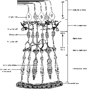
Figure 21.2The rods and cones are toward the rear of the retina, which causes the nerve impulses to go in the opposite direction of the light. (LifeART©1989-2001, Lippincott Williams & Wilkins)
The rods are more sensitive to light, but they are only used in perceiving the world as black and white. As such, we cannot see color by moonlight, but without light we cannot see anything, regardless of how long we wait for our eyes to adjust. Cones, on the other hand, perceive color, but a great deal more light is needed. The central fovea has the sharpest vision because of the high number of cones there. There are, however, no rods there; because of this, you cannot see anything in the fovea at night, but you can see objects if you look slightly off-center.
You might assume that we perceive light at the inner edge of the retina, but this is not the case. Partly to protect the retina from bright light, the rods and cones are near the back of the retina, in an area called the photoreceptor layer. Just behind this layer is a pigment epithelium, which uses melanin to prevent the reflection of light inside the eye. Light rays open ion channels that stimulate the cells of the bipolar cells, which transmit the message to the ganglion cells on the surface of the retina, and then to the optic nerve.

Excerpted from The Complete Idiot's Guide to Anatomy and Physiology © 2004 by Michael J. Vieira Lazaroff. All rights reserved including the right of reproduction in whole or in part in any form. Used by arrangement with Alpha Books, a member of Penguin Group (USA) Inc.
To order this book direct from the publisher, visit the Penguin USA website or call 1-800-253-6476. You can also purchase this book at Amazon.com and Barnes & Noble.
Trending
Here are the facts and trivia that people are buzzing about.